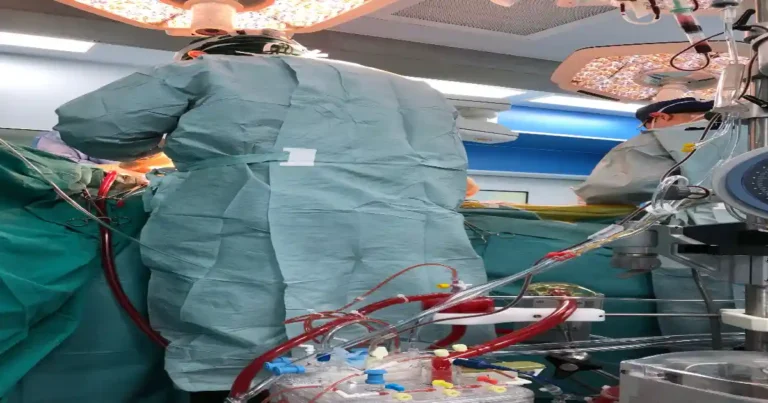
The occurrence of an empty venous reservoir during cardiopulmonary bypass (CPB) is a serious perfusion emergency that demands immediate recognition and intervention. In this blog post, we will explore a case study of an 83-year-old patient undergoing emergency mitral valve repair, where impaired venous return led to a critical drop in oxygenator blood levels. We will discuss the causes, solutions, and key takeaways for perfusionists and cardiac surgery teams. Additionally, we will delve into preventive measures, advanced troubleshooting techniques, and cutting-edge research related to venous drainage complications in cardiac surgery.
Case Overview
A critically ill 83-year-old obese female was admitted to the ICU with acute shortness of breath. Initial evaluation revealed severe mitral insufficiency due to a flail posterior leaflet. Following a diagnostic cardiac catheterization, the patient experienced sudden hypotension, tachycardia, and respiratory distress, requiring urgent surgical intervention.
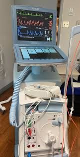
Upon initiation of cardiopulmonary bypass, the perfusionist noted a rapidly falling blood level in the oxygenator, despite adequate arterial return. Additionally, the right atrium became significantly distended, indicating impaired venous drainage.
Root Cause Analysis
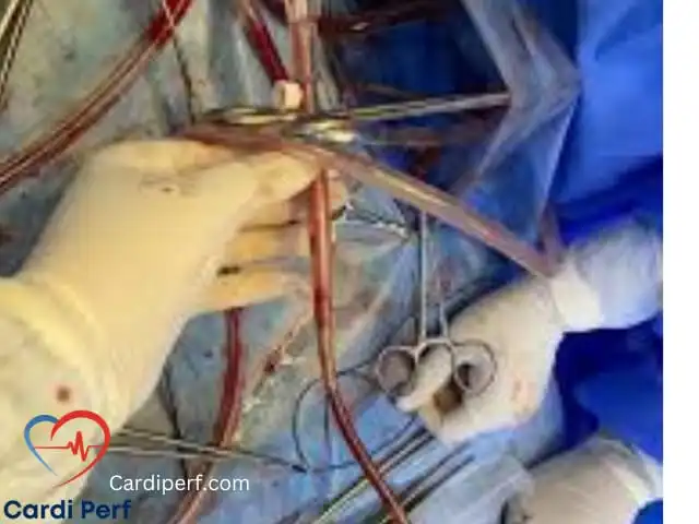
After verifying there were no kinks, clamps, or malpositioning of the two-stage venous cannula, the surgical team palpated the vena cava and noted an unusual spongy consistency. Upon exchanging the two-stage cannula for separate superior vena cava (SVC) and inferior vena cava (IVC) cannulas, they discovered multiple large organized thrombi obstructing venous return. These thrombi likely originated from the patient’s pelvic and deep femoral veins, resulting in pulmonary emboli and circulatory instability prior to surgery.
Immediate Solution and Intervention
- Exchange the Two-Stage Cannula – The two-stage venous cannula was replaced with separate SVC and IVC cannulas, connected via a Y-connector to restore flow.
- Remove Obstructing Thrombi – Large embolic thrombi (10 cm long) were extracted, allowing for full decompression of the right atrium and venous reservoir stabilization.
- Resume Full Flow on CPB – With improved venous return, the surgical team proceeded with mitral valve repair.
Potential Causes of Empty Venous Reservoir
Several factors may contribute to impaired venous return during CPB, including:
Mechanical Obstruction
- Thrombi or emboli within the venous system
- Malpositioned venous cannula obstructing flow
- Compression of major venous structures (e.g., by pericardial effusion or tumor)
Venous Cannula Issues
- Kinking of venous drainage lines
- Airlocks in venous tubing
- Clamps inadvertently left on venous lines
Physiological Considerations
- Hypovolemia leading to inadequate venous return
- High right atrial pressure reducing drainage efficiency
- Venous dilation causing improper cannula positioning
Advanced Troubleshooting Techniques
- Continuous Venous Pressure Monitoring – Maintain a constant vigilance on venous pressures to detect abnormalities early.
- Ultrasound-Guided Cannula Placement – Utilize intraoperative ultrasound to ensure optimal positioning of the venous cannula.
- Real-Time Transesophageal Echocardiography (TEE) – Use TEE to confirm right atrial decompression and detect any venous obstruction.
- Optimized Circuit Design – Employ venous reservoirs with enhanced flow dynamics to minimize the risk of airlocks.
- Patient Positioning Adjustments – Slight repositioning of the patient can sometimes improve venous return, especially in obese individuals.
Preventive Measures for Perfusionists and Cardiac Teams
- Preoperative Risk Stratification – Identify patients at high risk for venous thromboembolism and plan accordingly.
- Adequate Anticoagulation Management – Ensure optimal dosing of heparin to reduce clot formation risk.
- Regular Flushing of Venous Cannula – Prevent stagnation and clotting in venous lines by maintaining continuous low-flow perfusion.
- Routine System Checks – Conduct frequent inspections of the CPB circuit to identify potential obstructions before initiation.
- Improved Cannula Designs – Use newer-generation multi-stage venous cannulas designed to optimize flow and reduce clot formation.
Recent Research and Innovations
1. Advanced Cannulation Techniques
Recent studies suggest that the use of bicaval dual-stage cannulation significantly reduces the risk of venous obstruction compared to traditional two-stage cannulation. Hybrid cannulation strategies combining percutaneous and surgical approaches have also shown promise in high-risk patients.
2. Thromboprophylaxis Strategies
Ongoing research explores the use of novel anticoagulants, such as direct thrombin inhibitors, in CPB patients to reduce thrombus formation without excessive bleeding risk.
3. Computational Fluid Dynamics in CPB
Advanced modeling techniques now allow for the simulation of venous drainage patterns, helping surgeons anticipate and correct flow disturbances preemptively.
Key Lessons for Perfusionists
Understanding the potential causes of an empty venous reservoir is crucial for safe perfusion management. Here are key takeaways:
- Recognize the Signs Early: Monitor oxygenator levels, venous drainage, and right heart distension.
- Assess High-Risk Patients: Patients with a history of deep vein thrombosis (DVT), obesity, and prolonged hypotension may be at greater risk of venous embolism.
- Be Ready for Cannulation Adjustments: If venous return is impaired, consider switching to separate SVC and IVC cannulation.
- Monitor Venous Line Pressure: Elevated pressures may indicate obstruction due to thrombus, malposition, or extrinsic compression.
- Confirm No Kinks or Airlocks: Ensure smooth gravity drainage and avoid inadvertent clamps or air entry into the venous line.
- Optimize Anticoagulation Strategies: Preoperative and intraoperative anticoagulation protocols should be tailored for high-risk embolic patients.
FAQs
1. What is an empty venous reservoir?
An empty venous reservoir occurs when there is insufficient blood return from the patient’s venous system to the CPB circuit, leading to reduced perfusion and potential hemodynamic instability.
2. How can perfusionists prevent venous drainage obstruction?
Perfusionists can prevent venous drainage issues by ensuring proper anticoagulation, confirming correct cannula placement, and monitoring venous pressures throughout the procedure.
3. What role does transesophageal echocardiography (TEE) play in detecting venous return issues?
TEE provides real-time imaging of the heart and great vessels, allowing the surgical team to identify and correct venous drainage problems promptly.
4. How does hypovolemia contribute to venous return issues?
Hypovolemia reduces central venous pressure, making it more difficult for blood to drain effectively into the CPB circuit, leading to reservoir depletion.
5. Can venous return be improved by patient positioning?
Yes, minor adjustments in patient positioning, such as Trendelenburg positioning, can sometimes enhance venous return and improve CPB efficiency.
Conclusion
An empty venous reservoir is a high-stakes emergency that requires rapid assessment and decisive action. Stay connected with CardiPerf.com for more in-depth insights and the latest updates in perfusion science and cardiac surgery.