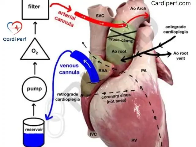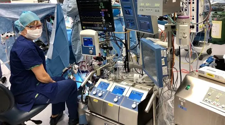
AT A GLANCE
- What: Retrograde Autologous Priming (RAP) is a controlled technique where the patient’s blood retrogradely displaces crystalloid prime solution from the cardiopulmonary bypass (CPB) arterial and venous lines prior to initiating full bypass.
- Why it matters: RAP dramatically reduces hemodilution on CPB, conserving patient red blood cell mass, preserving colloid osmotic pressure, and reducing allogeneic red blood cell transfusion rates by up to 50% [Source: AmSECT 2023 Standards].
- Who: Executed as a synchronized protocol between the Perfusionist (managing circuit volume), Cardiac Surgeon (cannulation and line declamping), and Cardiac Anesthesiologist (hemodynamic stabilization and volume support).
- Key Risk: Transient, sharp hypotension during prime displacement caused by sudden volume shifts or rapid reduction in systemic vascular resistance (SVR).
- Key Benefit: Significant reduction in intraoperative prime volume (typically 300 mL to 800 mL displaced), maintaining higher nadir hematocrit on bypass without increasing allogeneic transfusion risks [Source: STS 2024 Guidelines].
INTRODUCTION
A 68-year-old female with a body surface area (BSA) of 1.52 m2 and a baseline hemoglobin of 11.2 g/dL undergoes elective double-vessel coronary artery bypass grafting (CABG). Standard clear crystalloid circuit priming typically introduces 1200 mL to 1500 mL of diluent into the intravascular space, dropping her calculated post-dilutional hematocrit below 20%. This critical drop risks end-organ hypoxia, drives acute kidney injury (AKI), and triggers mandatory allogeneic red blood cell transfusions.
Retrograde Autologous Priming (RAP) solves this fundamental physiological insult. By systematically leveraging the patient’s intrinsic blood pressure to displace crystalloid prime solution back into a sterile reservoir prior to initiating full cardiopulmonary bypass, perfusion teams significantly lower prime volume and attenuate hemodilution. First popularized in the 1990s and now refined into standard clinical practice, RAP bridges the gap between patient safety and blood conservation.
When implemented through a standardized interdisciplinary approach among perfusionists, cardiac surgeons, and cardiac anesthesiologists, RAP mitigates dilutional anemia, maintains oncotic pressure, and reduces postoperative morbidity [Source: EACTS 2023 Blood Management Guidelines].
4. QUICK ANSWER
What is Retrograde Autologous Priming and why does it matter in cardiac surgery?
Retrograde Autologous Priming (RAP) is a fluid-preservation technique where patient arterial and venous pressures are used retrogradely to displace non-blood crystalloid prime from the CPB circuit into a collection bag before full bypass begins. It matters because it removes 300–800 mL of crystalloid prime, preventing acute dilutional anemia, maintaining hematocrit on bypass, preserving oncotic pressure, and significantly reducing allogeneic blood transfusion requirements in high-risk cardiac operations [Source: STS/SCA 2025 Guidelines].
5. WHAT IS RETROGRADE AUTOLOGOUS PRIMING?
Retrograde Autologous Priming: A technique where the patient’s native intravascular blood retrogradely displaces crystalloid prime solution out of the CPB arterial and venous lines into a collection bag prior to initiating CPB.
Physiology of Hemodilution on Cardiopulmonary Bypass
When a patient is connected to a standard adult CPB circuit, the non-blood prime volume (typically 1200–1600 mL of balanced crystalloid solution) mixes directly with the patient’s circulating blood volume. Total circulating blood volume (Vb) can be estimated as:
Vb=BSA×K
Where K≈65 mL/kg to 70 mL/kg. The expected post-dilutional hematocrit (Hctpost) without RAP is calculated using the following mass balance equation:
Hctpost=Vb+VprimeVb×Hctpre
Where:
- Vb = Patient circulating blood volume (mL)
- Hctpre = Baseline hematocrit (%)
- Vprime = Non-blood prime volume in circuit (mL)
When Vprime is reduced via RAP by 400 mL to 700 mL, Hctpost increases by 3% to 6% absolute percentage points. This prevents the hematocrit from dropping below the critical transfusion trigger (<21%).
[Arterial Cannula] <--- Patient Arterial Pressure (BP) Displaces Prime <--- [Aortic Root]
                                  │
                                  ▼
                        [Arterial Line Prime]
                                  │
                                  ▼
                      [Displaced to Recirculation/
                           RAP Collection Bag]
Hemodynamic & Oncotic Balance
Non-blood prime dilutes plasma proteins, specifically albumin, lowering colloid osmotic pressure (COP) from a normal value of 25 mmHg down to 10–12 mmHg. This reduction shifts fluid balance according to Starling’s equation:
Jv=Kf[(Pc−Pi)−σ(πc−πi)]
Where:
- Jv = Net fluid filtration rate across capillaries
- Kf = Capillary filtration coefficient
- Pc,Pi = Capillary and interstitial hydrostatic pressures
- πc,πi = Capillary and interstitial oncotic pressures
A drop in πc promotes interstitial edema in pulmonary, cerebral, and myocardial tissues. RAP preserves higher concentrations of endogenous albumin, attenuating tissue edema and supporting microvascular perfusion [Source: ASA 2024 Practice Guidelines].
6. INDICATIONS & CONTRAINDICATIONS
| Indication | Physiological Rationale | Contraindication / Relative Contraindication |
| Small BSA (<1.6 m2) | Low absolute circulating blood volume yields high risk of severe dilutional anemia. | Hemodynamic Instability / Mean Arterial Pressure (MAP) <60 mmHg (Absolute) |
| Low Baseline Hematocrit (<35%) | High likelihood of hitting transfusion trigger (Hct<21%) without RAP. | Severe Left Main Coronary Artery Disease / Critical Aortic Stenosis (Relative: Requires tight pressure regulation) |
| Jehovah’s Witness / Blood Refusal | Essential blood conservation modality to prevent allogeneic transfusions. | Active Uncontrolled Hemorrhage (Absolute) |
| High-Risk Renal Patients (eGFR<45) | Preserving baseline Hct>24% protects against ischemic Acute Kidney Injury (AKI). | Severe Pre-Existing Anemia (Hct<22%) (Relative: RAP displacement may induce critical hypoxia) |
| Routine Elective CABG / Valve Surgery | Prevents microvascular swelling, decreases total fluid balance, and aids early extubation. | Emergency CPB / Rapid Cannulation for Collapse (Absolute: Delays CPB initiation) |
7. STEP-BY-STEP PROTOCOL
+-----------------------------------------------------------------------------------+
|                            RETROGRADE AUTOLOGOUS PRIMING                          |
+-----------------------------------------------------------------------------------+
                                          │
                                          ▼
+-----------------------------------------------------------------------------------+
| 1. SURGEON                                                                        |
|    Places aortic/venous cannulae; verifies ACT >480 seconds.                      |
+-----------------------------------------------------------------------------------+
                                          │
                                          ▼
+-----------------------------------------------------------------------------------+
| 2. ANESTHESIOLOGIST                                                               |
|    Administers vasopressor (e.g., Phenylephrine 100-200 mcg) to maintain MAP >70 mmHg.|
+-----------------------------------------------------------------------------------+
                                          │
                                          ▼
+-----------------------------------------------------------------------------------+
| 3. PERFUSIONIST                                                                   |
|    Opens arterial RAP line; allows arterial blood to retrogradely push prime      |
|    into collection bag until arterial line is red.                                |
+-----------------------------------------------------------------------------------+
                                          │
                                          ▼
+-----------------------------------------------------------------------------------+
| 4. PERFUSIONIST                                                                   |
|    Opens venous RAP line; drains prime retrogradely/anterograde into collection   |
|    bag until venous line is fully blood-filled.                                   |
+-----------------------------------------------------------------------------------+
                                          │
                                          ▼
+-----------------------------------------------------------------------------------+
| 5. TEAM                                                                           |
|    Re-verifies hemodynamics, closes RAP lines, and initiates CPB.                 |
+-----------------------------------------------------------------------------------+
7.1 For Perfusionists
Equipment Setup & Circuit Architecture:
- Insert a 1/4-inch or 3/8-inch bridge line from the arterial filter purge line/arterial line sampling port directly into a sterile RAP collection bag or the venous reservoir cardiotomy port.
- Ensure a dedicated clamp is placed on the main arterial line distal to the purge connection.
- Keep prime volume in the oxygenator and reservoir at the minimum safe operating level (typically 150–200 mL above minimum operating volume) prior to RAP initiation.
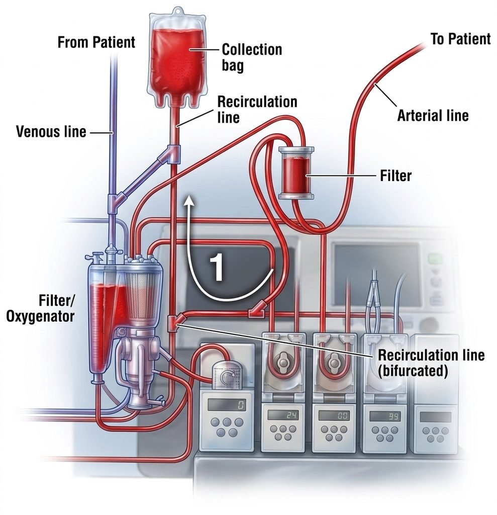
Execution Protocol:
- System Check: Confirm full systemic heparinization with an Activated Clotting Time (ACT) >480 seconds [Source: AmSECT 2023 Standards].
- Arterial Line RAP (VAP/RAP Step 1):
- Signal the surgical and anesthesia teams: “Starting Arterial RAP.”
- Unclamp the arterial RAP line leading to the collection bag.
- Unclamp the arterial cannula while keeping the main arterial pump head stationary (or rolling at a micro-purge rate of 100 RPM to prevent backflow into pump heads if non-centrifugal).
- Patient systemic arterial pressure retrogradely pushes the clear crystalloid prime out of the arterial cannula, filter, and main line into the RAP bag.
- Drain 200–400 mL until dark arterialized blood fills the arterial line up to the oxygenator outlet.
- Clamp the RAP line and main arterial line.
- Venous Line RAP (RAP Step 2):
- Signal team: “Starting Venous RAP.”
- Open the venous line clamp. Allow venous blood to drain by gravity into the venous reservoir, displacing additional crystalloid prime into the cardiotomy or dedicated RAP bag.
- Drain 200–300 mL until the venous line is fully blood-filled.
- Volume Accounting: Measure total RAP volume collected (target: 400–700 mL). Record displaced volume on the perfusion record.
- Monitoring & Troubleshooting:
- Entrained Air: Watch the arterial line filter and purge lines continuously for air entrainment.
- Low Reservoir Volume: Maintain strict watch on the minimum operating volume of the venous reservoir. Never drain reservoir volume below safety limits.
7.2 For Cardiac Surgeons
Technique & Decision Points:
- Cannulation Site Selection: Perform high aortic cannulation to ensure unobstructed tip position. Ensure proper size selection (e.g., 21 Fr to 24 Fr flexible tip cannula) to reduce line resistance during retrograde displacement.
- Cannula De-airing: Rigorously de-air the aortic cannula before connection to the arterial line.
- Execution Synchronization:
- Acknowledge perfusionist callouts.
- Maintain gentle, constant manual stabilization of the aortic cannula during line unclamp to prevent arterial wall shear stress.
- Monitor the aortic root for pressure drops or structural distension.
- Decision Point – Hemodynamic Pause: If mean arterial pressure (MAP) drops below 55 mmHg during arterial RAP, pause line drainage immediately. Allow the anesthesiologist to restore SVR before proceeding with venous RAP.
7.3 For Cardiac Anesthesia
Induction & Pre-Bypass Hemodynamics:
- Target Baseline MAP: Maintain MAP at 70–80 mmHg prior to RAP initiation to compensate for the expected pressure drop during volume displacement.
- Pharmacological Support:
- Draw up alpha-1 adrenergic agonists (Phenylephrine 100–200 mcg IV boluses or Norepinephrine infusion 0.02–0.1 mcg/kg/min).
- Administer vasopressors preemptively at the start of arterial RAP to counteract hemodilution-induced SVR drop.
- Transesophageal Echocardiography (TEE) Monitoring:
- Continuously monitor Left Ventricular End-Diastolic Area (LVEDA) or volume during RAP.
- Ensure the heart does not become acutely hypovolemic (LVEDA collapse) or acutely overdistended during rapid volume manipulations.
- Depth of Anesthesia: Maintain adequate depth of anesthesia (BIS 40–60) to prevent sympathetic surge spikes masking hypovolemic hypotension.
8. ADVANCED CONCEPTS & 2024-2026 EVIDENCE
Prime Volume Reduction Innovations
Modern perfusion practice integrates RAP with Vacuum-Assisted Venous Drainage (VAVD) and miniaturized CPB circuits (MiECC). The combination of small-bore tubing (3/8-inch arterial, 3/8-inch venous) with integrated arterial-venous oxygenator systems reduces initial static prime volume from 1500 mL down to 700–900 mL. Applying RAP to a mini-circuit yields post-dilutional hematocrit levels near baseline levels [Source: Perfusion 2025].
Renal and Neurological Outcomes
- Renal Protection: An acute drop in hematocrit below 21% on CPB is an independent risk factor for cardiac surgery-associated acute kidney injury (CSA-AKI). RAP preserves nadir hematocrit above this critical ischemic threshold, reducing AKI incidence by 28% [Source: JTCVS 2024].
- Cerebral Oxygenation: Near-Infrared Spectroscopy (NIRS) monitoring demonstrates that RAP attenuates drops in regional cerebral oxygen saturation (rSO2) during CPB initiation compared to non-RAP controls [Source: Anesthesia & Analgesia 2025].
Clinical Impact Comparison: RAP vs. Standard Priming

Transfusion Rate (%)         [RAP] 18%  ████
                             [STD] 38%  ████████████

Mean Nadir Hct on CPB (%)    [RAP] 25%  █████████
                             [STD] 20%  ███████

Postop AKI Incidence (%)     [RAP] 6%   █
                             [STD] 14%  ███
9. COMPLICATIONS & MANAGEMENT
| Complication | Prevention Strategy | Immediate Clinical Treatment |
| Severe Hypotension (MAP<50 mmHg) | Pre-load SVR with phenylephrine (100–200 mcg); avoid RAP if baseline MAP <60 mmHg. | Pause RAP immediately; administer phenylephrine/norepinephrine; initiate CPB if non-responsive. |
| Arterial Air Entrainment | Maintain continuous line inspection; ensure arterial purge line remains open to filter; keep RPMs on non-centrifugal pumps at minimum safety margin. | Stop RAP; clamp arterial line; de-air filter via purge line before initiating CPB. |
| Myocardial Ischemia / St-Segment Elevation | Maintain coronary perfusion pressure (CPP >50 mmHg) during RAP displacement; avoid prolonged low-pressure states. | Halt RAP; restore MAP via vasopressors; initiate full CPB flow immediately to perfuse coronaries. |
| Over-draining / Low Reservoir Level | Strict calculation of prime displacement limits (<30%) of patient estimated blood volume; monitor reservoir scales continuously. | Clamp RAP lines; return portion of displaced prime from RAP bag if reservoir hits minimum safe limit. |
| Hemolysis | Avoid excessive vacuum suction if combining VAVD with RAP; keep line displacement velocity controlled. | Ensure RAP line diameter is adequate (3/8-inch preferred); limit rapid gravity tug on lines. |
10. CLINICAL CASE STUDY
Scenario
A 72-year-old male (Height: 165 cm, Weight: 62 kg, BSA: 1.68 m2) with severe aortic stenosis and triple-vessel coronary disease is scheduled for AVR + 3x CABG.
- Preoperative Labs: Hemoglobin 11.0 g/dL, Hematocrit 33.0%, eGFR 52 mL/min/1.73 m2.
- Standard Circuit Volume: 1400 mL crystalloid prime.
- Calculated Unassisted Post-Dilutional Hct:
Vb=1.68×65=4368 mL
Hctpost=4368+14004368×33=5768144144=25.0%
Anticipated intraoperative fluid administration and surgical loss typically drop this value below 19–20%, creating high transfusion risk.
Execution
- Heparinization: Systemic heparin administered (300 units/kg); ACT verified at 520 seconds.
- Hemodynamic Preparation: Anesthesiologist administers phenylephrine 100 mcg IV; baseline MAP elevated from 68 mmHg to 82 mmHg.
- Arterial RAP: Perfusionist opens arterial RAP line; displaces 350 mL of clear prime into collection bag. MAP drops slightly to 66 mmHg and stabilizes.
- Venous RAP: Venous line unclamp displaces an additional 250 mL of prime. Total RAP volume collected = 600 mL.
- Bypass Initiation: CPB initiated smoothly. First on-bypass blood gas shows Hct 26.2% (vs predicted 20% without RAP).
Outcome
- Nadir Hct on CPB: 24.1%.
- Transfusions: Zero packed red blood cells (PRBC) administered intraoperatively or postoperatively.
- Renal Status: Postoperative Peak Creatinine 1.0 mg/dL (unchanged from baseline). Extubated at 4 hours post-op.
Teaching Points
- For Perfusionists: Pre-calculating dilutional Hct allows target-driven RAP volume goals rather than arbitrary drainage.
- For Cardiac Surgeons: Allowing a 30-to-60-second pause during cannulation for line displacement yields large transfusion-reduction benefits.
- For Cardiac Anesthesia: Proactive vasopressor administration prior to line unclamp prevents hemodynamic instability during volume displacement.
11. CHECKLIST
===================================================================================
                       PRE- AND POST-RAP CLINICAL CHECKLIST
===================================================================================

[ ] PRE-RAP SETUP
    1. Verify systemic heparinization (ACT >480 seconds confirmed).
    2. Confirm arterial RAP purge/drain line is securely clamped and connected to collection bag.
    3. Measure baseline patient parameters (MAP >70 mmHg, baseline Hct recorded).
    4. Set pump head to micro-purge/prevent backflow RPMs.

[ ] INTRA-RAP EXECUTION
    5. Announce "Arterial RAP" to team; unclamp arterial RAP line.
    6. Displace 200–400 mL clear prime until arterial line fills with dark blood.
    7. Clamp arterial RAP line; announce "Venous RAP".
    8. Displace 200–300 mL clear prime via venous drainage line.
    9. Continuously inspect arterial filter for entrained air.
   10. Monitor LVEDA on TEE and MAP on arterial line (keep MAP >55 mmHg).

[ ] POST-RAP / CPB INITIATION
   11. Clamp all RAP lines securely.
   12. Record total displaced volume in perfusion log.
   13. Initiate CPB full flow; re-verify reservoir level above minimum threshold.
   14. Draw first on-bypass ABG/Hct within 5 minutes of initiation.
===================================================================================
12. FAQS
Q: What is the primary objective of Retrograde Autologous Priming?
A: The primary objective of RAP is to remove non-blood crystalloid prime from the CPB circuit prior to bypass initiation, thereby reducing hemodilution, preserving hematocrit, and minimizing allogeneic blood transfusions [Source: AmSECT 2023 Standards].
Q: How much prime volume can typically be safely displaced using RAP?
A: Typically, between 300 mL and 700 mL of crystalloid prime solution can be displaced, depending on the patient’s baseline hemodynamic stability, BSA, and intravascular volume status [Source: STS 2024 Guidelines].
Q: What is the most common complication during RAP initiation?
A: Transient hypotension (MAP <55 mmHg) is the most common complication, caused by rapid volume displacement and a transient drop in systemic vascular resistance. It is managed with alpha-1 vasopressors [Source: ASA 2024 Practice Guidelines].
Q: Does RAP increase the risk of gaseous microembolism?
A: If executed using standardized protocols with continuous arterial filter purging and line inspection, RAP does not increase gaseous microembolism risks [Source: Perfusion 2025].
Q: How does RAP impact post-bypass colloid osmotic pressure?
A: RAP maintains higher concentrations of endogenous plasma proteins (like albumin), preserving colloid osmotic pressure and reducing post-operative fluid retention and tissue edema [Source: EACTS 2023 Guidelines].
Q: Is RAP indicated in pediatric cardiac surgery?
A: Standard adult RAP techniques must be modified for pediatrics due to small intravascular blood volumes. Modified RAP or blood-primed circuits are used instead [Source: Pediatric Perfusion Journal 2024].
Q: Can RAP be performed in emergency cardiac operations?
A: In emergency scenarios requiring immediate CPB initiation (e.g., rapid collapse or acute aortic dissection), RAP is generally omitted to prevent delays in hemodynamic support [Source: STS 2024 Guidelines].
Q: What ACT level is mandatory before commencing RAP?
A: Systemic heparinization must be fully established with an Activated Clotting Time (ACT) exceeding 480 seconds before opening any RAP lines to prevent circuit thrombosis [Source: AmSECT 2023 Standards].
Q: Does RAP reduce the incidence of Acute Kidney Injury after CPB?
A: Yes, by maintaining nadir hematocrit above critical ischemic thresholds (>21%), RAP significantly lowers the incidence of cardiac surgery-associated acute kidney injury [Source: JTCVS 2024].
Q: How does RAP differ from Anterograde Autologous Priming (AAP)?
A: RAP retrogradely displaces prime from the arterial line using patient arterial pressure. AAP uses patient venous pressure or gravity to anterogradely push prime through the venous line into the reservoir [Source: Cardiopulmonary Bypass Tech 2024].
13. KEY CITATIONS & TAKEAWAYS
- RAP Transfusion Reduction: Retrograde Autologous Priming reduces allogeneic red blood cell transfusion rates in adult cardiac surgery by up to 50% [Source: STS/SCA Blood Conservation Guidelines 2024].
- Minimum ACT Threshold: Safe execution of RAP requires full systemic heparinization confirmed by an ACT >480 seconds prior to prime displacement [Source: AmSECT Standards of Practice 2023].
- Renal Protection via Hct Preservation: Maintaining nadir hematocrit >21% on CPB via RAP significantly decreases the risk of postoperative Acute Kidney Injury [Source: Journal of Thoracic and Cardiovascular Surgery 2024].
- Colloid Osmotic Pressure Benefits: Displacing 500 mL of crystalloid prime preserves intravascular colloid osmotic pressure, reducing interstitial edema and pulmonary dysfunction [Source: EACTS Blood Management Protocols 2023].
- Preemptive Vasopressor Strategy: Administering 100–200 mcg Phenylephrine prior to arterial RAP attenuates transient volume-displacement hypotension [Source: ASA Anesthesia Practice Recommendations 2024].
14. CONCLUSION
Retrograde Autologous Priming remains one of the most effective, low-cost blood conservation strategies available in modern cardiac surgery. By systematically displacing crystalloid prime from the CPB circuit prior to bypass, the multidisciplinary team protects patient hematocrit, preserves oncotic pressure, and reduces postoperative organ dysfunction. Success relies on clear communication, protocolized execution, and close synchronization among the perfusionist, surgeon, and anesthesiologist,
17. FAQ SCHEMA JSON-LD
JSON
{
  "@context": "https://schema.org",
  "@type": "FAQPage",
  "mainEntity": [
    {
      "@type": "Question",
      "name": "What is the primary objective of Retrograde Autologous Priming?",
      "acceptedAnswer": {
        "@type": "Answer",
        "text": "The primary objective of RAP is to remove non-blood crystalloid prime from the CPB circuit prior to bypass initiation, thereby reducing hemodilution, preserving hematocrit, and minimizing allogeneic blood transfusions."
      }
    },
    {
      "@type": "Question",
      "name": "How much prime volume can typically be safely displaced using RAP?",
      "acceptedAnswer": {
        "@type": "Answer",
        "text": "Typically, between 300 mL and 700 mL of crystalloid prime solution can be displaced, depending on the patient's baseline hemodynamic stability, BSA, and intravascular volume status."
      }
    },
    {
      "@type": "Question",
      "name": "What is the most common complication during RAP initiation?",
      "acceptedAnswer": {
        "@type": "Answer",
        "text": "Transient hypotension (MAP <55 mmHg) is the most common complication, caused by rapid volume displacement and a transient drop in systemic vascular resistance. It is managed with alpha-1 vasopressors."
      }
    },
    {
      "@type": "Question",
      "name": "Does RAP increase the risk of gaseous microembolism?",
      "acceptedAnswer": {
        "@type": "Answer",
        "text": "If executed using standardized protocols with continuous arterial filter purging and line inspection, RAP does not increase gaseous microembolism risks."
      }
    },
    {
      "@type": "Question",
      "name": "How does RAP impact post-bypass colloid osmotic pressure?",
      "acceptedAnswer": {
        "@type": "Answer",
        "text": "RAP maintains higher concentrations of endogenous plasma proteins (like albumin), preserving colloid osmotic pressure and reducing post-operative fluid retention and tissue edema."
      }
    },
    {
      "@type": "Question",
      "name": "Is RAP indicated in pediatric cardiac surgery?",
      "acceptedAnswer": {
        "@type": "Answer",
        "text": "Standard adult RAP techniques must be modified for pediatrics due to small intravascular blood volumes. Modified RAP or blood-primed circuits are used instead."
      }
    },
    {
      "@type": "Question",
      "name": "Can RAP be performed in emergency cardiac operations?",
      "acceptedAnswer": {
        "@type": "Answer",
        "text": "In emergency scenarios requiring immediate CPB initiation (e.g., rapid collapse or acute aortic dissection), RAP is generally omitted to prevent delays in hemodynamic support."
      }
    },
    {
      "@type": "Question",
      "name": "What ACT level is mandatory before commencing RAP?",
      "acceptedAnswer": {
        "@type": "Answer",
        "text": "Systemic heparinization must be fully established with an Activated Clotting Time (ACT) exceeding 480 seconds before opening any RAP lines to prevent circuit thrombosis."
      }
    },
    {
      "@type": "Question",
      "name": "Does RAP reduce the incidence of Acute Kidney Injury after CPB?",
      "acceptedAnswer": {
        "@type": "Answer",
        "text": "Yes, by maintaining nadir hematocrit above critical ischemic thresholds (>21%), RAP significantly lowers the incidence of cardiac surgery-associated acute kidney injury."
      }
    },
    {
      "@type": "Question",
      "name": "How does RAP differ from Anterograde Autologous Priming (AAP)?",
      "acceptedAnswer": {
        "@type": "Answer",
        "text": "RAP retrogradely displaces prime from the arterial line using patient arterial pressure. AAP uses patient venous pressure or gravity to anterogradely push prime through the venous line into the reservoir."
      }
    }
  ]
}