A struck aortic valve is a life-threatening emergency where a mechanical prosthetic valve becomes obstructed, typically due to thrombus formation, pannus overgrowth, or structural failure. This obstruction severely restricts blood flow, leading to acute heart failure, cardiogenic shock, or sudden cardiac arrest. The condition demands immediate surgical intervention, as delays can result in irreversible organ damage or death.
The pathophysiology of a stuck valve varies—thrombus-related obstructions often arise from inadequate anticoagulation, while pannus (fibrous tissue growth) develops gradually. Perfusionists must recognize these differences, as they influence surgical and bypass strategies. For instance, thrombotic obstructions may require thrombolysis before surgery, whereas pannus-related cases need direct valve replacement.
Table of Contents
The Perfusionist’s Critical Role
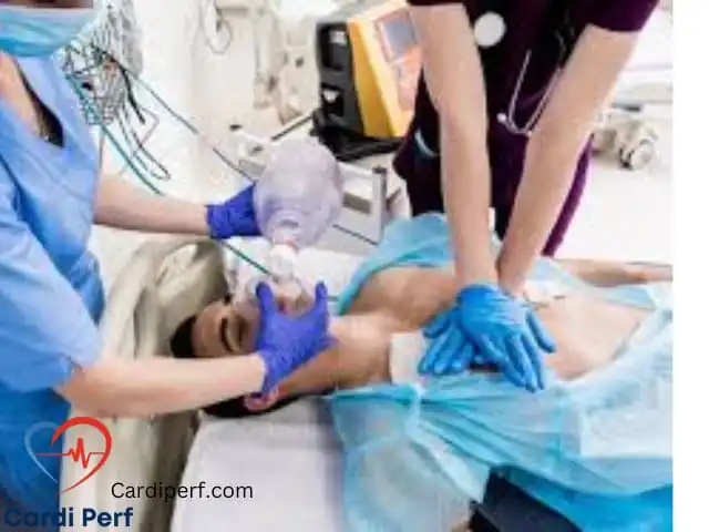
Perfusionists are central to managing struck aortic valve emergencies, ensuring the rapid and safe initiation of cardiopulmonary bypass (CPB). Their expertise in hemodynamic management, circuit priming, and anticoagulation monitoring directly impacts patient survival. Unlike routine cardiac surgeries, struck valve cases require ultra-fast CPB deployment, often under unstable conditions.
Additionally, perfusionists must anticipate complications such as massive air embolism, bleeding, or poor venous return. Their ability to troubleshoot in real-time—adjusting flows, administering blood products, or switching to backup circuits—can mean the difference between life and death. Effective communication with surgeons and anesthesiologists is equally crucial, as coordinated efforts minimize delays and errors.

Struck Valve as a Redo Surgery
Most struck aortic valve cases are redo surgeries, meaning the patient has undergone prior cardiac procedures. This introduces complexities such as adhesions, altered anatomy, and higher bleeding risks. Unlike first-time sternotomies, re-entry carries a risk of accidental injury to the heart, great vessels, or existing grafts.
Perfusionists must prepare for alternative cannulation strategies, such as femoral or axillary artery access, in case central cannulation is unsafe. They should also be ready for sudden hemodynamic collapse during sternal re-entry, requiring immediate CPB initiation. Given these challenges, preoperative planning—including reviewing past surgical notes and imaging—is essential for optimizing outcomes.
Pre-Operative Perfusion Considerations
Hemodynamic Assessment and Support
Before initiating CPB, perfusionists must assess the patient’s hemodynamic status. A stuck aortic valve often presents with severe hypotension, pulmonary edema, or cardiogenic shock, necessitating aggressive pharmacologic support. Inotropes like epinephrine or milrinone may be required to maintain cardiac output, while vasopressors such as norepinephrine help sustain perfusion pressure.
Additionally, echocardiography plays a key role in confirming valve obstruction and evaluating ventricular function. Perfusionists should collaborate closely with the anesthesia team to optimize pre-bypass hemodynamics, ensuring the patient is stable enough for CPB initiation. In extreme cases, mechanical circulatory support (e.g., ECMO) may be needed as a bridge to surgery.
Equipment Preparation and Readiness
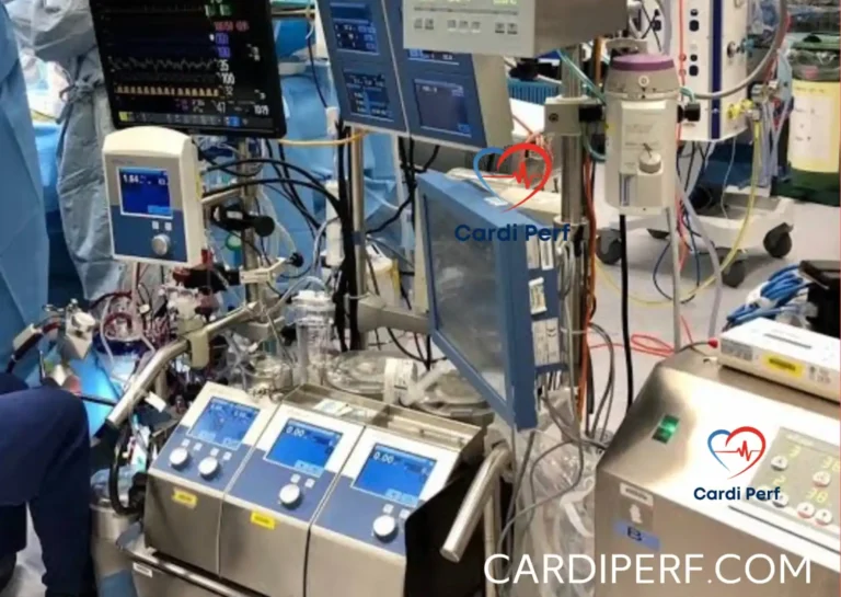
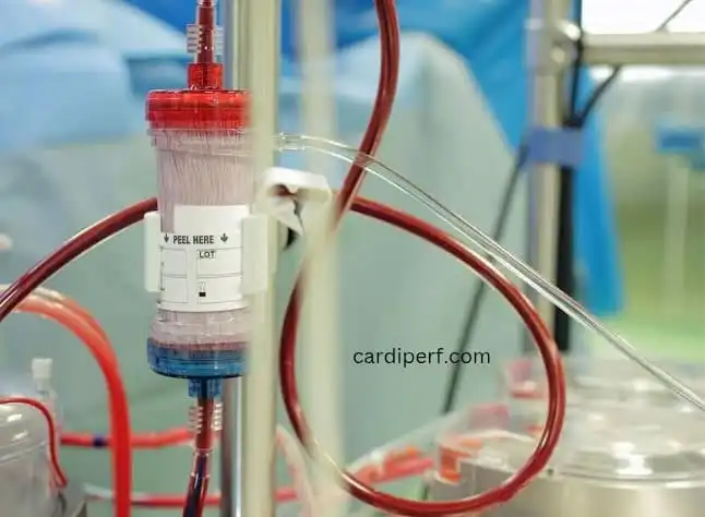
Given the urgency of struck aortic valve cases, perfusionists must have all equipment prepped and primed before the patient arrives. The CPB circuit should be checked for leaks, proper oxygenation, and adequate flow capacity. Backup components—such as an extra oxygenator, tubing pack, or centrifugal pump—should be readily available in case of circuit failure.
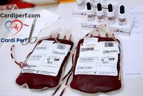
Blood product management is another critical aspect. Since redo surgeries carry a higher bleeding risk, perfusionists should ensure crossmatched blood, platelets, and fresh frozen plasma (FFP) are on standby. A cell saver system should also be primed to minimize allogeneic blood transfusions.

Femoral Cannulation Considerations
In redo sternotomies, femoral cannulation is often the safest approach due to mediastinal adhesions and scar tissue. However, this method has its own challenges. Ultrasound-guided vessel localization is essential to avoid complications like arterial dissection or misplaced cannulation.
Perfusionists must also monitor limb perfusion during femoral bypass, as prolonged cannulation can lead to ischemia or compartment syndrome. If femoral access is compromised, axillary or central aortic cannulation may be necessary.

Femoral Cannula Size and Dilator Sets
Selecting the correct cannula size is crucial for adequate flow. For adults, femoral arterial cannulas (18-24 Fr) and venous cannulas (21-25 Fr) are typically used. Larger cannulas improve flow but may increase vascular injury risk.
Dilator sets facilitate smooth cannula insertion, especially in calcified or tortuous vessels. Perfusionists should ensure these are available and compatible with the chosen cannulas.
Intra-Operative Perfusion Management
Cardiopulmonary Bypass Initiation
In struck aortic valve cases, CPB must be initiated rapidly but controllably. Sudden transitions can cause cerebral hypoperfusion or ventricular distension. Perfusionists should gradually increase pump flow while monitoring arterial pressure and venous saturation.
If femoral cannulation is used, retrograde flow may require higher pressures. Close monitoring of radial and femoral arterial line waveforms helps detect malperfusion.
Maintaining Perfusion During Valve Replacement
Once on bypass, perfusionists must optimize flow rates (typically 2.2-2.5 L/min/m²) while maintaining mean arterial pressure (MAP) >60 mmHg. Temperature management is also key—mild hypothermia (32-34°C) may be used to protect organs during prolonged cases.
Blood gas and electrolyte monitoring should be frequent (every 15-30 min) to prevent acidosis or electrolyte imbalances. Hemoglobin levels must be kept >7 g/dL to ensure adequate oxygen delivery.
Specific Considerations for Redo Sternotomy
Redo sternotomies increase the risk of catastrophic hemorrhage during dissection. Perfusionists should be prepared for sudden blood loss, ensuring rapid transfusion capabilities and cell saver use.
If adhesions prevent safe aortic clamping, deep hypothermic circulatory arrest (DHCA) may be required. Perfusionists must then manage cooling/rewarming phases carefully to avoid neurologic injury.
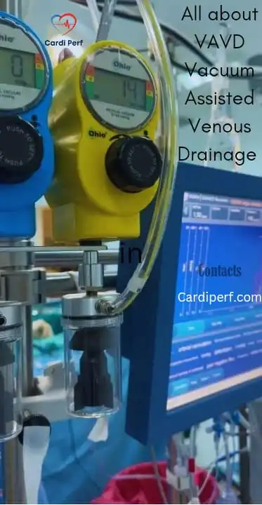
Vacuum-Assisted Venous Drainage (VAVD)
In cases of poor venous return, VAVD enhances drainage by applying controlled negative pressure (typically -20 to -40 mmHg). However, excessive suction can cause foaming or hemolysis, requiring close monitoring.
Weaning from CPB
Weaning should be gradual, with careful assessment of cardiac contractility, rhythm, and filling pressures. If the heart struggles, inotropic support or intra-aortic balloon pump (IABP) may be needed.
Post-CPB, bleeding management is critical. Antifibrinolytics (e.g., tranexamic acid) and factor concentrates may be administered to reduce transfusion needs.
Post-Operative Perfusion Considerations
Hemodynamic Monitoring in ICU
After surgery, continuous hemodynamic monitoring is essential. Echocardiography, lactate levels, and mixed venous saturation (SvO₂) help assess recovery.
Anticoagulation and Antiplatelet Therapy
Since mechanical valves require lifelong anticoagulation, perfusionists should ensure smooth transition from heparin to warfarin. ACT and INR monitoring must be rigorous to prevent thrombosis or bleeding.
Long-Term Perfusion Implications
CPB can cause renal injury, neurocognitive deficits, or systemic inflammation. Perfusionists should advocate for organ-protective strategies, such as goal-directed perfusion and minimized hemodilution.
Case Studies and Lessons Learned
Case Study: 16-Year Post-AVR Struck Aortic Valve Emergency
A 58-year-old male with a 16-year-old mechanical AVR presented with sudden hypotension and pulmonary edema. TEE confirmed valve thrombosis, prompting emergency surgery.
Key Actions:
- Femoral cannulation due to unstable hemodynamics.
- VAVD optimized venous drainage.
- Cell saver reduced transfusion needs.
Lessons:
- Preoperative imaging could have identified early pannus formation.
- Team drills improved response time.
Frequently Asked Questions (FAQs)
- Q: When is femoral cannulation preferred over central cannulation in a struck aortic valve redo?
- A: Femoral cannulation is preferred when severe adhesions or anatomical difficulties make central cannulation high-risk.
- Q: What are the key considerations for selecting femoral cannula sizes?
- A: Vessel size, patient BSA, and desired flow rates are the primary factors.
- Q: How does VAVD improve venous drainage?
- A: VAVD creates a negative pressure gradient, enhancing venous return to the CPB circuit.
- Q: What are the risks associated with femoral cannulation?
- A: Risks include vessel injury, dissection, hematoma, and distal limb ischemia.
- Q: What is the perfusionist’s role during the valve explant stage?
- A: Maintaining hemodynamic stability and providing optimal flow during a potentially unstable period.
- Q: How is ACT managed during and after struck aortic valve surgery?
- A: ACT is maintained at a high level during CPB, and post-op anticoagulation is managed according to the patient’s risk profile.
- Q: What is the importance of a dilator set during femoral cannulation?
- A: Dilator sets allow for progressive dilation of the vessel, minimizing trauma during cannula insertion.
- Q: How does a struck aortic valve affect pre-op hemodynamics?
- A: It can cause severe hemodynamic instability due to obstruction of blood flow.
- Q: What temperature management strategies are used during these cases?
- A: Mild to moderate hypothermia is often used for organ protection.
- Q: What is the perfusionist’s role in the post-operative period?
- A: Monitoring hemodynamic stability, and renal function, and communicating with the ICU team.
Conclusion
Managing struck aortic valves demands expert perfusion strategies, rapid decision-making, and seamless teamwork. By mastering femoral cannulation, VAVD, and emergency CPB protocols, perfusionists play a life-saving role in these high-risk cases.
Future advancements—such as miniaturized bypass systems and real-time perfusion analytics—could further improve outcomes. Until then, preparedness, communication, and precision remain the cornerstones of success.