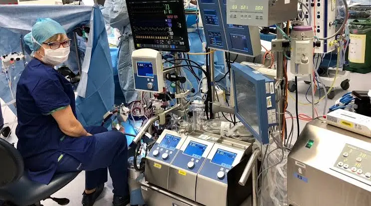
Clinical Perfusion in Cardiac Surgery: Sustaining Life Through Extracorporeal Circulation
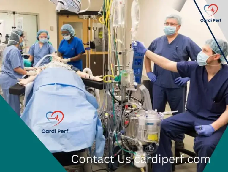
Clinical perfusion in cardiac surgery is the specialized medical practice of temporarily taking over a patient’s heart and lung functions using a cardiopulmonary bypass (CPB) machine. Operated by a certified clinical perfusionist, this extracorporeal circulation system drains deoxygenated blood, adds oxygen, removes carbon dioxide, regulates temperature, and pumps oxygenated blood back into the body so surgeons can operate safely on a still, bloodless heart.
While cardiac surgeons and anesthesiologists are widely recognized members of the operating room, clinical perfusionists serve as the physiological lifeline during complex open-heart procedures. Understanding how clinical perfusion works—from hemodynamic management to myocardial protection—reveals the engineering and medical precision required to make modern heart surgery possible.
What Is Clinical Perfusion and What Does a Perfusionist Do?
A clinical perfusionist is a highly trained healthcare professional who operates the heart-lung machine and manages a patient’s physiological status during cardiac and thoracic surgeries. Beyond running mechanical pumps, the perfusionist acts as the patient’s temporary circulatory and respiratory system, continuously adjusting blood flow, oxygenation, acid-base balance, and anticoagulation in real time.
According to clinical standards outlined by organizations such as the American Society of ExtraCorporeal Technology (AmSECT), a perfusionist’s core intraoperative responsibilities include:
- Extracorporeal Circulation Management: Assembling, priming, and operating the cardiopulmonary bypass (CPB) circuit to maintain systemic blood flow.
- Hemodynamic and Gas Exchange Control: Regulating arterial blood pressure, venous oxygen saturation (SvO₂), and arterial blood gases (pO₂ and pCO₂) to prevent end-organ hypoxia.
- Myocardial Protection (Cardioplegia): Administering specialized potassium-rich cardioplegic solutions to safely arrest the heart and preserve heart muscle tissue during surgical repair.
- Anticoagulation and Blood Conservation: Managing systemic heparinization to prevent clotting in the circuit, neutralizing heparin with protamine afterward, and operating autotransfusion (cell-saver) devices to recycle the patient’s own red blood cells.
- Thermoregulation: Cooling the patient’s blood (induced hypothermia) to lower metabolic oxygen demand during complex repairs and safely rewarming them before weaning off bypass.
How Does Cardiopulmonary Bypass Work During Open-Heart Surgery?
Cardiopulmonary bypass works by diverting blood away from the heart and lungs through venous cannulas into an external circuit, where a mechanical pump and artificial membrane oxygenator oxygenate the blood before returning it to the aorta. This closed-loop system isolates the heart from the rest of the circulatory system while keeping the brain, kidneys, and vital organs fully perfused.
The Step-by-Step Clinical Perfusion Workflow
- Systemic Anticoagulation: Before connecting the patient to the circuit, high-dose heparin is administered. The perfusionist verifies an Activated Clotting Time (ACT) typically exceeding 400–480 seconds to ensure blood will not clot upon contact with artificial tubing surfaces.
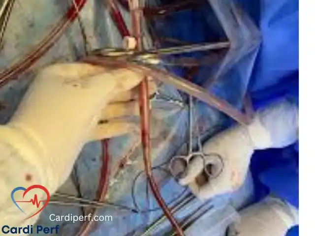
- Cannulation: The cardiac surgeon inserts drainage cannulas into the right atrium or superior and inferior vena cavae, and an arterial return cannula into the ascending aorta or femoral artery.
- Initiation of Bypass: Once safe ACT levels are confirmed, clamps are removed and venous blood drains by gravity or vacuum assistance into the venous reservoir.
- Gas Exchange and Filtration: A centrifugal or roller pump pushes blood through a heat exchanger and a membrane oxygenator, where oxygen diffuses into the blood and carbon dioxide is swept away. An arterial line filter traps micro-emboli or air bubbles before the blood re-enters the patient.
- Aortic Cross-Clamping and Cardioplegia: The surgeon places a cross-clamp across the aorta and delivers cardioplegia into the coronary arteries, stopping electrical and mechanical heart activity so delicate valve or bypass grafting work can proceed.
- Rewarming and Weaning: After surgical repair is complete, the perfusionist gradually rewarms the blood to normothermia (37°C), corrects electrolyte levels, and slowly reduces pump flow as the patient’s native heart resumes pumping.
Key Physiological Parameters Monitored in Cardiac Perfusion
To prevent postoperative complications such as acute kidney injury (AKI), stroke, or systemic inflammatory response syndrome (SIRS), perfusionists adhere to strict physiological targets. Modern clinical practice relies heavily on Goal-Directed Perfusion (GDP), an evidence-based approach focused on maintaining adequate oxygen delivery (DO₂) rather than relying solely on fixed flow formulas.
| Physiological Parameter | Typical Clinical Target Range | Primary Clinical Purpose |
|---|---|---|
| Cardiac Index (Flow Rate) | 2.2 – 2.4 L/min/m² (normothermic) | Ensures systemic tissue perfusion matches metabolic demand. |
| Mean Arterial Pressure (MAP) | 50 – 80 mmHg (patient-dependent) | Maintains cerebral and renal autoregulation during bypass. |
| Indexed Oxygen Delivery (DO₂i) | > 280 – 300 mL/min/m² | Prevents cellular hypoxia and reduces postoperative acute kidney injury. |
| Venous Oxygen Saturation (SvO₂) | 70% – 80% | Indicates the balance between systemic oxygen delivery and consumption. |
| Activated Clotting Time (ACT) | > 400 – 480 seconds | Prevents thrombus formation inside the extracorporeal circuit. |
| Arterial Blood Temperature | 28°C – 37°C (procedure-specific) | Lowers cellular metabolic rate during complex or prolonged repairs. |
Types of Perfusion Technologies Beyond Standard Bypass
While conventional cardiopulmonary bypass is the backbone of coronary artery bypass grafting (CABG) and valve replacement, clinical perfusion encompasses several specialized modalities tailored to patient acuity.
1. Minimally Invasive Extracorporeal Circulation (MiECC)
Conventional CPB circuits require significant priming volumes of crystalloid solution, which can dilute red blood cells (hemodilution) and trigger inflammatory pathways when blood contacts air and foreign surfaces. MiECC systems utilize compact, closed, biocompatible-coated circuits without an open venous reservoir. Studies featured in the National Library of Medicine (PMC) highlight that minimized circuits reduce hemodilution, lower inflammatory markers, and decrease the need for allogeneic red blood cell transfusions.
2. Extracorporeal Membrane Oxygenation (ECMO)
Unlike standard CPB, which supports a patient for a few hours in the operating room, ECMO provides prolonged cardiac and respiratory life support in the intensive care unit for days or weeks.
- Veno-Arterial (VA) ECMO: Supports both heart and lung function in patients suffering from cardiogenic shock or post-cardiotomy heart failure.
- Veno-Venous (VV) ECMO: Supports lung function exclusively in severe respiratory failure, such as acute respiratory distress syndrome (ARDS).
3. Deep Hypothermic Circulatory Arrest (DHCA)
For complex surgeries involving the aortic arch or congenital heart defects, surgeons may need a completely cannula-free, bloodless field. The perfusionist cools the patient’s core temperature to between 18°C and 20°C, temporarily halting all blood circulation while protecting the brain via selective antegrade or retrograde cerebral perfusion.
4. Mechanical Circulatory Support (MCS) and Organ Perfusion
Perfusionists also manage ventricular assist devices (VADs), intra-aortic balloon pumps (IABPs), and Ex Vivo Heart Perfusion (EVHP) systems. EVHP pumps warm, oxygenated, nutrient-rich blood through donor hearts outside the body, extending safe transport times and expanding the viable donor pool for heart transplantation.
Safety Protocols and Risk Mitigation in Clinical Perfusion
Modern cardiac perfusion incorporates redundant mechanical safeguards and continuous electronic monitoring to make catastrophic circuit complications exceedingly rare. Because even brief interruptions in oxygenated blood flow can cause irreversible neurological injury, perfusion protocols follow rigorous international safety checklists.
- Air Embolism Prevention: Ultrasonic bubble detectors and venous reservoir level sensors automatically trigger alarms or clamp arterial lines if air enters the circuit or blood levels drop dangerously low.
- Pressure Monitoring: Continuous line pressure sensors detect kinks, cannula malposition, or aortic dissection risks before excessive shear stress damages red blood cells (hemolysis).
- Inline Blood Gas Monitoring: Optical sensors continuously track arterial and venous oxygen saturation, hemoglobin, hematocrit, and electrolytes without waiting for intermittent lab draws.
- Backup Hardware: Every heart-lung machine is equipped with battery backups and hand cranks to ensure uninterrupted circulation in the event of a power or motor failure.
The Future of Clinical Perfusion: AI, Data Integration, and Biocompatibility
The next decade of clinical perfusion is shifting toward automated data integration, artificial intelligence-assisted decision support, and advanced biomimetic circuit coatings.
- Real-Time Predictive Analytics: Modern perfusion consoles integrate electronic medical records with live hemodynamic telemetry, alerting perfusionists to subtle trends in oxygen delivery (DO₂) or lactate buildup before organ stress occurs.
- Advanced Surface Coatings: Heparin-bonded and phosphorylcholine-coated tubing mimic the natural endothelial lining of human blood vessels, further minimizing platelet activation and postoperative coagulopathy.
- Personalized Pediatric Perfusion: Ultra-low prime volume circuits and miniaturized oxygenators allow neonatal and pediatric heart surgeries to be performed with minimal or zero donor blood transfusions, significantly reducing immunologic complications in infants.
Frequently Asked Questions (FAQ)
What is the difference between a cardiac perfusionist and a cardiac anesthesiologist?
A cardiac anesthesiologist is a medical doctor (MD/DO) responsible for inducing anesthesia, managing pain, monitoring transesophageal echocardiography (TEE), and overseeing overall perioperative medical care. A clinical perfusionist is a specialized allied health professional who operates the heart-lung machine, manages extracorporeal blood flow, and administers cardioplegia while the heart is stopped.
How long can a patient safely stay on a cardiopulmonary bypass machine?
Most routine open-heart surgeries require 1 to 3 hours on cardiopulmonary bypass. While complex aortic or multi-valve surgeries can safely extend beyond 4 hours with careful temperature and oxygen delivery management, longer bypass durations carry higher risks of systemic inflammation and bleeding, making efficient surgical and perfusion coordination critical.
What qualifications are required to become a clinical perfusionist?
Educational pathways vary globally. In the United States, candidates typically earn a bachelor’s degree in science followed by a 2-year accredited Master’s program in cardiovascular perfusion and board certification through the American Board of Cardiovascular Perfusion (ABCP). Similar accredited BSc or MSc clinical training programs exist in the UK, Europe, Australia, and Pakistan.
For more details visit us at cardiperf.com