Extracorporeal Membrane Oxygenation (ECMO) is a vital intervention that provides temporary heart and lung support to critically ill patients. The process of ECMO cannulation, which involves inserting cannulas into blood vessels to connect the patient to an ECMO circuit, is intricate and requires a multidisciplinary team approach. This guide delves into the step-by-step process of ECMO cannulation for Veno-Arterial (V-A) and Veno-Venous (V-V) ECMO, highlighting considerations for pediatric cases, advanced imaging techniques, and equipment details. It is tailored for healthcare professionals, including anesthesiologists, cardiologists, cardiac surgeons, pediatric cardiac surgeons, perfusionists, ECMO specialists, intensivists, and specialized nurses.
Table of Contents
ECMO Cannulation Types
Veno-Arterial (V-A) ECMO
- Purpose: Supports both cardiac and respiratory functions by bypassing the heart and lungs.
- Cannulation Sites:
- Arterial: Femoral artery, axillary artery, or ascending aorta.
- Venous: Right internal jugular vein or femoral vein.
- Indications: Cardiogenic shock, cardiac arrest, or failure to wean from cardiopulmonary bypass.
Veno-Venous (V-V) ECMO
- Purpose: Provides respiratory support while the heart continues to function.
- Cannulation Sites:
- Central: Right internal jugular vein (drainage) and right femoral vein (return).
- Peripheral: Dual femoral veins or a single-site double-lumen cannula in the right internal jugular vein.
- Indications: Acute respiratory distress syndrome (ARDS), severe pneumonia, or refractory hypoxemia.

Step-by-Step Guide to ECMO Cannulation
Patient Assessment and Preparation
- Clinical Evaluation: Assess the patient’s hemodynamic status, organ function, and coagulation profile.
- Informed Consent: Obtain consent from the patient or their family.
- Preparation:
- Administer appropriate sedation and analgesia.
- Secure venous access for medications and fluids.
- Monitor coagulation parameters and adjust anticoagulation therapy as needed.
Equipment and Personnel
Essential Equipment:
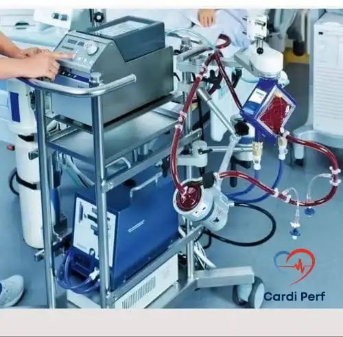
- ECMO circuit (e.g., Rotaflow, Cardio-Help) with oxygenator, pump, and heat exchanger.
- Cannulas:
- Adults: 17-21 French (arterial), 23-27 French (venous).
- Pediatrics: Size based on Body Surface Area (BSA) and ELSO guidelines.
- Wires (flexible, stiff, and Super Stiff), dilators, and needles.
- Imaging devices (fluoroscopy, ultrasound).
- Hemostatic agents and anticoagulation therapy (e.g., heparin).
- Surgical instruments (scalpels, sutures, clamps).
Multidisciplinary Team:
- ECMO specialists, intensivists, cardiothoracic surgeons, anesthesiologists, perfusionists, nurses, and imaging technicians.
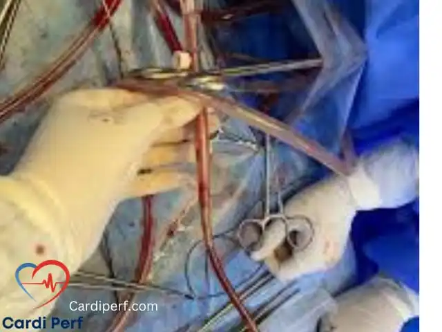
Cannulation Process
V-A ECMO Cannulation:
- Arterial Cannulation:
- Insert a flexible guidewire into the femoral or axillary artery.
- Use imaging (fluoroscopy or TEE) to confirm placement.
- Dilate the vessel and insert the arterial cannula.
- Secure the cannula with sutures.
- Venous Cannulation:
- Insert the guidewire into the femoral or internal jugular vein.
- Confirm placement using ultrasound or TEE.
- Insert the venous cannula and secure.
V-V ECMO Cannulation:
- Single-Site Approach:
- Use a double-lumen cannula (e.g., Avalon) in the internal jugular vein.
- Confirm positioning with TEE for optimal drainage and return flow.
- Dual-Site Approach:
- Insert drainage and return cannulas into separate veins (e.g., femoral and jugular).
- Confirm placement and flow direction with imaging.
Circuit Connection and Initiation
- Connect the cannulas to the ECMO circuit.
- Prime the circuit and initiate blood flow.
- Adjust flow rates based on patient needs.
- Continuously monitor for complications (e.g., bleeding, thrombosis, or hemolysis).
Post-Cannulation Management
- Monitor ECMO settings and patient parameters.
- Administer anticoagulation therapy to prevent thrombosis.
- Provide hemodynamic and respiratory support.
- Initiate weaning protocols as clinically indicated.
Case-Based Scenario
Scenario:
A 52-year-old male presents in cardiogenic shock following an acute myocardial infarction. Despite maximal medical therapy, his condition deteriorates, necessitating V-A ECMO initiation.
Challenges:
- Obesity (BMI 34) complicates vascular access.
- Severe coagulopathy increases the risk of bleeding.
Solution:
- Use femoral arterial and venous cannulation.
- Begin with a flexible wire and transition to a Super Stiff wire for stability.
- Employ TEE to confirm wire placement and assess for complications.
- Securely insert and fix 19 French arterial and 25 French venous cannulas.
Outcome:
The patient stabilizes with improved organ perfusion and is successfully weaned from ECMO after five days.
Special Considerations
- Imaging Guidance: Use TEE or TTE to confirm wire placement, cannula positioning, and flow adequacy.
- Wire Selection: Flexible wires are ideal for initial access, while Super Stiff wires provide stability in challenging cases.
- Complications: Be vigilant for vessel perforation, thrombosis, air embolism, and infection.
- Pediatric Cases: Choose cannula sizes and configurations based on BSA and follow ELSO guidelines.
- ECMO Systems: Familiarize with Rotaflow, Cardio-Help, and other systems to optimize circuit management.
Conclusion
ECMO cannulation is a critical procedure that requires precision, preparation, and a collaborative approach. By following this guide and leveraging advanced imaging and equipment, healthcare teams can enhance patient outcomes and minimize risks.
SEO Considerations
- Primary Keywords: ECMO cannulation, V-A ECMO, V-V ECMO, pediatric ECMO, TEE, TTE.
- Secondary Keywords: Rotaflow, Cardio-Help, ECMO initiation, multidisciplinary ECMO teams.
- Meta Description: “Step-by-step ECMO cannulation guide for V-A and V-V ECMO, tailored for healthcare teams. Includes pediatric insights, imaging tips, and case studies.”
Disclaimer: This guide is for educational purposes only and should not replace professional medical consultation. Always refer to institutional protocols for clinical decisions.
Step-by-Step Guide to ECMO Cannulation
Patient Assessment and Preparation
- Evaluate the patient’s clinical condition, including hemodynamic stability, organ function, and coagulation status.
- Obtain informed consent from the patient or their family.
- Prepare the patient with adequate sedation and analgesia.
- Ensure sufficient venous access for medications and fluids.
- Monitor coagulation parameters and adjust anticoagulation therapy as needed.
Equipment and Personnel
Equipment:
- ECMO circuit (e.g., Rotaflow, Cardio-Help) and components (oxygenator, pump, heater-exchanger).
- Cannulas of appropriate size based on patient needs (refer to site selection).
- Wires (flexible, stiff, and Super Stiff).
- Dilators, needles, surgical instruments, imaging equipment (fluoroscopy, ultrasound).
- Hemostatic agents and anticoagulation therapy.
- Monitoring equipment for blood pressure, oxygen saturation, and other vital parameters.
Personnel:
- ECMO specialists, intensivists, cardiothoracic surgeons, anesthesiologists, nurses, and imaging technicians.
Cannulation Site Selection and Cannula Size
Veno-Arterial (V-A) ECMO:
- Arterial Cannulation:
- Sites: Ascending aorta, femoral artery.
- Cannula Size:
- Adults: 17-21 French
- Pediatrics: Based on Body Surface Area (BSA) and ELSO (Extracorporeal Life Support Organization) guidelines.
- Venous Cannulation:
- Sites: Right internal jugular vein, right femoral vein.
- Cannula Size:
- Adults: 23-27 French
- Pediatrics: Refer to BSA and ELSO guidelines.
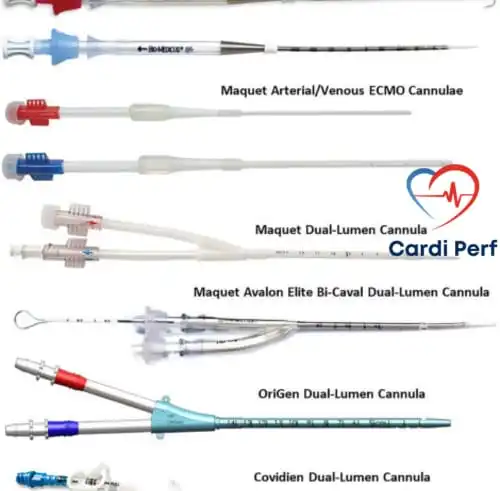
Veno-Venous (V-V) ECMO:
- Venous Cannulation:
- Sites:
- Central: Right internal jugular vein (drainage) and right femoral vein (return).
- Peripheral: Both femoral veins.
- Cannula Size:
- Adults: 17-21 French (drainage), 19-23 French (return).
- Pediatrics: Follow BSA and ELSO guidelines.
Pediatric ECMO:
- Arterial Cannulation:
- Sites: Femoral artery, axillary artery, ascending aorta (for neonates).
- Venous Cannulation:
- Sites: Right internal jugular vein, femoral vein, jugular vein (for neonates).
- Cannula Size: Based on BSA and ELSO recommendations.
Cannulation Process
Aortic Cannulation (V-A ECMO):
- Access Site Selection: Ascending aorta or femoral artery.
- Wire Insertion:
- Start with a flexible wire and confirm placement using imaging (fluoroscopy or echocardiography).
- Cannulation:
- Advance the dilator and arterial cannula over the wire. Confirm placement and secure with sutures.
Venous Cannulation (V-A and V-V ECMO):
- Access Site Selection: Right internal jugular vein, right femoral vein, or external jugular vein.
- Wire Insertion:
- Use a flexible wire initially; switch to a Super Stiff wire if needed.
- Confirm placement with imaging.
- Cannulation:
- Advance the dilator and venous cannula over the wire, ensuring proper placement with imaging and securing with sutures.

ECMO Circuit Initiation
- Connect the patient to the ECMO circuit.
- Initiate blood flow and adjust flow rates as needed.
- Monitor for complications, such as bleeding, thrombosis, or organ dysfunction.
Post-Cannulation Management
- Monitor the patient closely and adjust ECMO settings as required.
- Provide supportive care, including anticoagulation and infection prevention.
- Wean the patient from ECMO as clinically appropriate.
Case-Based Discussion
Scenario:
A 52-year-old male patient presents in cardiogenic shock following an acute myocardial infarction. Despite maximal medical therapy, his condition worsens, necessitating ECMO initiation.
Challenge: The patient has a BMI of 34, complicating vascular access and wire passage.
Solution:
- Femoral V-A ECMO is selected.
- A flexible wire is used initially, transitioning to a Super Stiff wire for stability. Resistance during wire passage is resolved with fluoroscopic guidance and gradual advancement.
- TEE confirms wire placement and rules out complications.
- Cannula sizes (19 French arterial and 25 French venous) are chosen to ensure optimal flow rates.
- Post-cannulation, the patient stabilizes with improved perfusion and organ function.
Outcome: The patient is successfully weaned from ECMO after five days, with significant recovery.
Additional Considerations
- Wire Visualization: Prevents vessel perforation and ensures smooth cannulation.
- Role of TTE and TEE:
- TEE offers superior visualization, particularly for venous wire placement.
- Both modalities assist in guiding cannula insertion and identifying complications.
- Complications:
- Be vigilant for vessel perforation, bleeding, thrombosis, and infection.
- ECMO Systems: Familiarize with systems like Rotaflow and Cardio-Help to optimize usage.
Conclusion
ECMO cannulation is a critical, multidisciplinary procedure requiring meticulous preparation and precision. By following these steps and leveraging imaging modalities, healthcare providers can improve patient outcomes and minimize complications. This guide serves as a valuable resource for teams involved in ECMO Cannulation. visit updated content at cardiperf.com.
Disclaimer: This guide is for educational purposes only and should not replace professional medical consultation. Always refer to institutional protocols for clinical decisions.