Positron Emission Tomography (PET)scan is an advanced molecular imaging technique that plays a crucial role in diagnosing and monitoring various diseases, including cancer, heart conditions, and neurological disorders. PETscan imaging provides detailed metabolic insights, offering advantages beyond conventional imaging methods like CT or MRI.
Table of Contents
How PET Imaging Works
PET imaging utilizes radioactive tracers (radiopharmaceuticals) injected into the bloodstream. These tracers emit positrons, which interact with electrons in the body, producing gamma rays. A PET scanner detects these gamma rays and creates detailed 3D images of metabolic activity in tissues and organs.
Key Steps in PET Imaging:
- Tracer Injection: The patient receives an intravenous (IV) injection of a radiotracer.
- Absorption Period: The tracer circulates and accumulates in target tissues.
- Imaging Process: The PET scanner captures metabolic activity and generates images.
- Data Analysis: Specialists interpret the scans to diagnose or monitor conditions.
Key Applications of PET Imaging
PET scans are widely used across multiple medical fields:
1. Cardiology: Identifying Heart Disease
- Detects coronary artery disease (CAD) by assessing blood flow.
- Identifies areas of the heart with reduced oxygen supply (ischemia).
- Helps evaluate heart function post-surgery or intervention.
2. Oncology: Cancer Detection and Monitoring
- Provides early detection of tumors before they become visible on other scans.
- Helps in staging cancer and determining its spread (metastasis).
- Monitors treatment response and recurrence.
3. Neurology: Diagnosing Brain Disorders
- Detects Parkinson’s disease and epilepsy-affected brain areas.
- Assesses brain tumors and strokes effectively.
- Identifies Alzheimer’s disease and dementia by mapping brain metabolism.
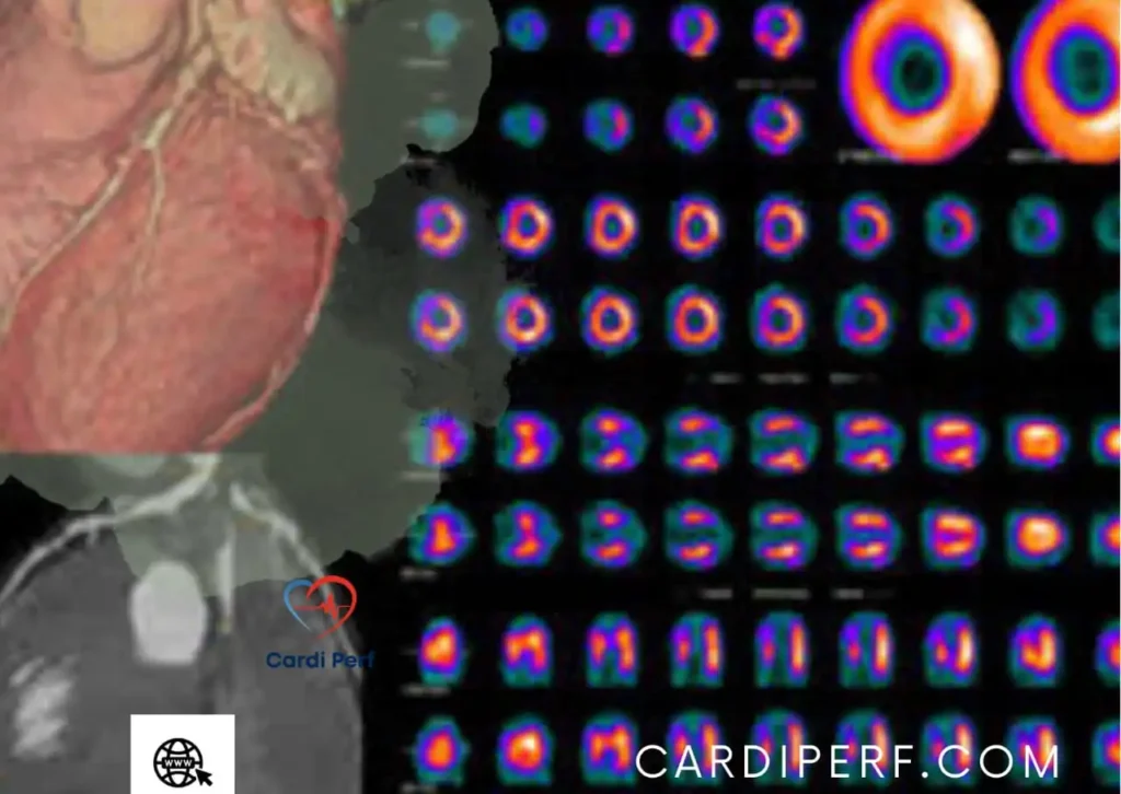
PET Imaging in Cardiology
PET imaging plays a crucial role in cardiology by providing highly accurate functional imaging of the heart. It helps in:
- Detecting Coronary Artery Disease (CAD): PET imaging can pinpoint regions with reduced blood flow and assess the severity of arterial blockages.
- Assessing Myocardial Viability: Determines if damaged heart tissue is still viable for treatment.
- Monitoring Post-Intervention Recovery: Helps track the success of treatments like angioplasty or bypass surgery.
- Evaluating Heart Failure: Identifies inflammation or metabolic dysfunction in heart muscles.
PET vs. Other Imaging Techniques
| Imaging Type | Function | Strengths | Limitations |
|---|---|---|---|
| PET Imaging | Functional imaging | Detects metabolic changes early | Exposure to radiation |
| CT Scan | Structural imaging | High-resolution anatomical details | Limited metabolic data |
| MRI Scan | Soft tissue imaging | No radiation, excellent tissue contrast | Expensive and time-consuming |
| X-ray | Basic structural imaging | Quick and cost-effective | Low detail in soft tissues |
PET imaging excels in detecting diseases at an early stage, often before structural changes occur.
Frequently Asked Questions (FAQs)
1. What is PET imaging used for?
PET imaging is used for diagnosing and monitoring cancer, heart disease, and neurological conditions by detecting metabolic activity in tissues.
2. How long does a PET scan take?
The entire process takes about 30 to 90 minutes, depending on the area being scanned.
3. Is PET imaging painful?
No, the scan itself is painless. The only discomfort might be from the IV injection of the radiotracer.
4. Are there any risks associated with PET imaging?
PET imaging involves low radiation exposure, but it’s generally safe. Pregnant women should avoid it unless necessary.
5. How do I prepare for PET imaging?
Avoid eating 6-12 hours before the scan and inform your doctor about any medications you are taking.
6. Can PET imaging detect all cancers?
PET imaging is highly effective in detecting many cancers, but some tumors may not absorb the tracer efficiently.
7. What is the difference between PET imaging and MRI?
PET imaging shows metabolic activity, while an MRI provides detailed structural images of soft tissues.
8. How often can I have PET imaging?
Doctors determine the frequency based on the condition being monitored. Repeated scans should be limited due to radiation exposure.
9. Do PET scans require contrast dye?
No, PET scans use radioactive tracers, not contrast dye, though some hybrid PET-CT scans may use both.
10. Is PET imaging better than a CT scan for heart disease?
Yes, PET imaging provides detailed blood flow and metabolic data, making it superior for detecting early heart disease.
Conclusion
PET imaging has revolutionized medical diagnostics, offering unparalleled insights into metabolic activity and disease progression. With applications ranging from cardiology and oncology to neurology, PET scanning remains a critical tool for early diagnosis, treatment planning, and patient care. As technology advances, PET imaging will continue to push the boundaries of medical diagnostics, ensuring better patient outcomes. For update about cardiac Perfusion Sciences visit us at cardiperf.com